I. Introduction: Navigating the Curve: The Role of Corneal Topography in Custom Contact Lens Fittings
Corneal topography is a sophisticated diagnostic tool that maps the surface of the cornea, the eye’s outermost layer. This non-invasive imaging technique provides detailed information about the shape and curvature of the cornea, which is crucial for diagnosing and treating various eye conditions. Its precision and ability to capture minute details make corneal topography indispensable in the realm of ophthalmology, especially in the fitting of specialized contact lenses.
The importance of corneal topography extends beyond basic eye examinations. It plays a pivotal role in custom contact lens fittings, particularly for individuals with unique corneal shapes or conditions that affect the cornea. By accurately mapping the cornea’s surface, corneal topography allows eye care professionals to design contact lenses that fit the individual’s eye perfectly, enhancing comfort and improving vision.
This technology is especially beneficial for patients with conditions such as keratoconus, post-surgical corneal changes, or other corneal deformities, where standard contact lenses may not provide adequate vision correction or comfort.
In essence, corneal topography serves as a foundation for personalized eye care, enabling the creation of contact lenses tailored to the specific needs of an individual’s eyes. This introduction will delve into the workings of corneal topography, its significance in diagnosing and treating eye conditions, and its critical role in custom contact lens fittings, setting the stage for a deeper exploration of this advanced technology and its benefits for those in need of specialized contact lenses.
II. Understanding Corneal Topography
Corneal topography is a highly detailed method used to map the surface of the cornea, the clear, dome-shaped surface that covers the front of the eye. This technology is crucial for assessing the cornea’s shape and curvature, which are vital for diagnosing eye conditions and designing contact lenses, especially for those with irregular corneas.
Technology Behind Corneal Topography
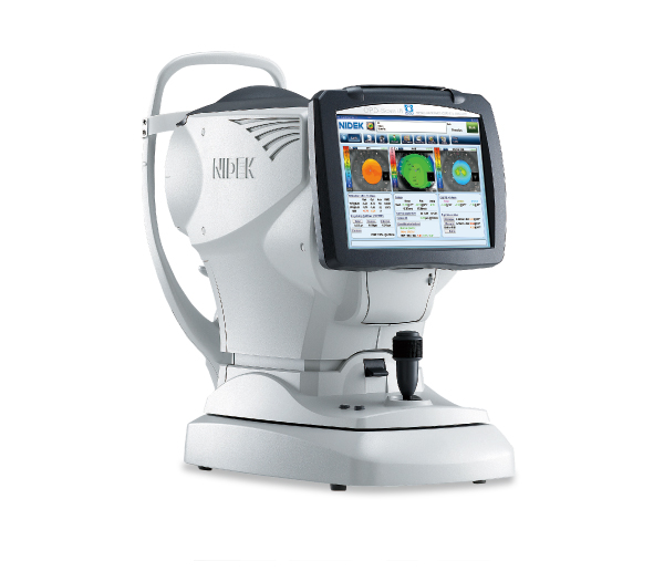
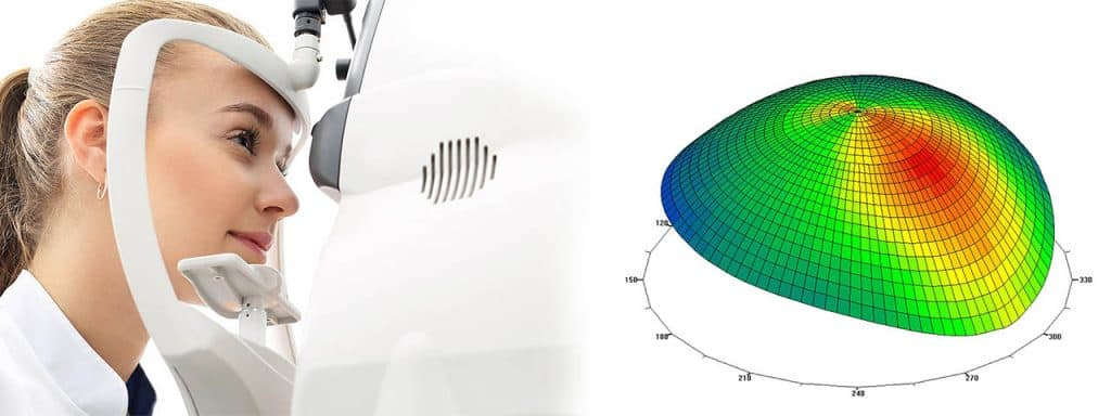
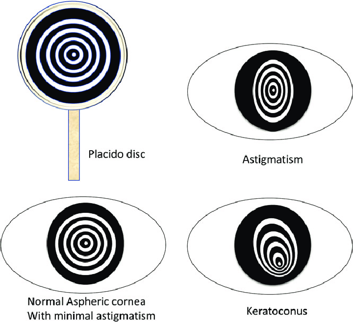
The primary technology used in corneal topography involves projecting a series of concentric rings, known as a Placido disc, onto the cornea and capturing the reflected image with a camera. This image is then analyzed to determine the curvature of the cornea across different zones.
Advanced systems like the Scheimpflug camera go further by providing a three-dimensional scan of the cornea, capturing both the anterior and posterior surfaces, which is essential for a comprehensive assessment [3].
Recent advancements have integrated optical coherence tomography (OCT) with corneal topography, allowing simultaneous corneal epithelial thickness mapping and topography from a single measurement. This integration enhances the detail and accuracy of the corneal maps produced, providing invaluable data for customizing contact lens fittings and other medical interventions [1].
Visuals and Diagrams
Visual aids in understanding corneal topography typically include diagrams of the Placido disc system, showing how the rings are reflected off the cornea and captured by the imaging system.
Additionally, screenshots or outputs from devices like the REVO NX or Pentacam illustrate the detailed maps produced. These maps highlight variations in corneal thickness and curvature, crucial for diagnosing conditions like keratoconus or planning surgical interventions [1][3].
Interpreting the Data
The data from corneal topography is presented in various forms, such as color-coded maps, numerical data, and graphical displays showing different corneal parameters like curvature, elevation, and thickness.
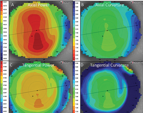
Each color or pattern in a topographic map corresponds to a specific range of curvature, with warmer colors typically indicating steeper areas and cooler colors representing flatter areas. This data is critical for identifying irregularities such as astigmatism or corneal ectasia, which are often precursors to more serious conditions [3].
The accuracy and reliability of these measurements are supported by high intraclass correlation coefficients (ICC), indicating excellent repeatability and reproducibility of the measurements, which is essential for monitoring changes over time or assessing the impact of treatments like corneal cross-linking in conditions like keratoconus [1][2].
In summary, corneal topography provides a detailed and accurate representation of the cornea’s surface, essential for diagnosing eye conditions, planning surgeries, and designing custom contact lenses. The technology’s ability to provide detailed, repeatable, and reliable data makes it an indispensable tool in modern ophthalmology.
References:
- [1] Sikorski, Bartosz L. “Simultaneous Corneal Topography and Epithelial Thickness Mapping from a Single Measurement Using Optical Coherence Tomography.” Journal of ophthalmology vol. 2022 7339306. 21 Apr. 2022, doi:10.1155/2022/7339306. https://www.ncbi.nlm.nih.gov/pmc/articles/PMC9050264/
- [2] Гальбинур, А П and П. И. Мусаев. “ANALYSIS OF CORNEAL TOPOGRAPHY DATA AT MEDIUM STAGES OF KERATOCONUS.” (2016).. https://www.semanticscholar.org/paper/2ab65b915991a2b2153977082cb4b97de8e1156b
- [3]Fan, Rachel et al. “Applications of corneal topography and tomography: a review.” Clinical & experimental ophthalmology vol. 46,2 (2018): 133-146. doi:10.1111/ceo.13136. https://pubmed.ncbi.nlm.nih.gov/29266624/
III. Who Needs Specialized Contact Lenses?
Specialized contact lenses are not just an alternative to standard eyewear; they are a necessity for individuals with certain ocular conditions that affect the cornea’s shape and function.
These conditions can significantly impair vision and, in some cases, standard contact lenses or glasses cannot provide adequate correction or comfort. Understanding who needs these specialized lenses is crucial for effective vision management.
Conditions Necessitating Specialized Contact Lenses
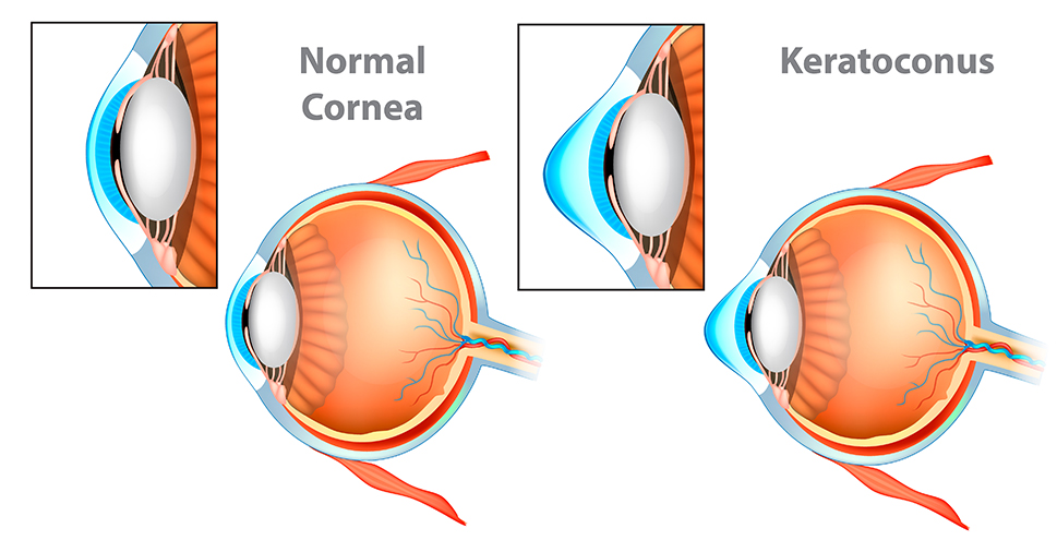
- Keratoconus is a progressive eye disorder in which the cornea gradually thins and begins to bulge outward into a cone-like shape, causing visual distortion.
- Traditional contact lenses often fail to accommodate the irregular corneal surface of keratoconus patients, making specialized lenses essential for vision correction [4][5][6][7].
- Post-surgical Corneal Changes: Surgical procedures on the eye, such as LASIK or corneal transplants, can alter the cornea’s shape. Patients may experience irregular astigmatism or other corneal irregularities post-surgery, necessitating custom-fitted lenses for optimal vision correction [4].
- Other Corneal Deformities and Irregularities: Conditions like pellucid marginal degeneration or significant astigmatism due to corneal scars or dystrophies also require specialized contact lenses. These lenses are designed to vault over the cornea, providing a smooth refractive surface that corrects vision more effectively than standard lenses [4][6].
Case Studies and Examples
- Keratoconus: A study involving 340 eyes of 254 keratoconus patients assessed the performance of corneal rigid gas-permeable (RGP) contact lens fitting software integrated with anterior segment optical coherence tomography (AS-OCT). The software’s ability to accurately predict the back optical zone radius (BOZR) for lenses tailored to various keratoconus morphologies demonstrates the importance of specialized fitting in managing this condition [4].
- Post-CXL Surgery: Another study focused on the mid-term effects of corneal collagen cross-linking (CXL) for progressive keratoconus and its impact on RGP contact lens fitting parameters. The findings highlighted changes in corneal topography post-CXL, which necessitated adjustments in contact lens fittings to accommodate the altered corneal shape [5].
These examples underscore the critical role of specialized contact lenses in managing complex corneal conditions. By providing tailored vision correction, these lenses can significantly improve the quality of life for individuals with challenging ocular irregularities.
References:
- [4] Itoi, Motohiro et al. “Corneal RGP Contact Lens Fitting Software for Keratoconus Built-In Anterior Segment Optical Coherence Tomography.” Eye & contact lens vol. 48,12 (2022): 503-508. doi:10.1097/ICL.0000000000000940. https://pubmed.ncbi.nlm.nih.gov/36223291/
- [5] Zhao, C P et al. [Zhonghua yan ke za zhi] Chinese journal of ophthalmology vol. 58,4 (2022): 272-278. doi:10.3760/cma.j.cn112142-20220129-00029. https://pubmed.ncbi.nlm.nih.gov/35391514/
- [6] Richter, Kathrin et al. “Contact Lens Fitting in Patients with Keratoconus – A Retrospective Assessment of 200 Patients.” “Verlauf der Kontaktlinsenanpassung bei Keratokonus – eine retrospektive Untersuchung bei 200 Patienten.” Klinische Monatsblatter fur Augenheilkunde vol. 239,9 (2022): 1155-1163. doi:10.1055/a-1526-9861. https://pubmed.ncbi.nlm.nih.gov/34731900/
- [7] Ortiz-Toquero, Sara et al. “Gas permeable contact lens fitting in keratoconus: Comparison of different guidelines to back optic zone radius calculations.” Indian journal of ophthalmology vol. 67,9 (2019): 1410-1416. doi:10.4103/ijo.IJO_1538_18. https://www.ncbi.nlm.nih.gov/pmc/articles/PMC6727727/
IV. The Role of Corneal Topography in Lens Fitting
Corneal topography has revolutionized the way eye care professionals approach contact lens fitting, particularly for rigid gas permeable (RGP) lenses and specialized conditions like keratoconus and post-surgical corneal changes. This section delves into how corneal topography guides the creation of custom contact lenses and the benefits it brings to both patients and practitioners.
Guiding Custom Contact Lens Creation
The precision of corneal topography allows for a detailed analysis of the cornea’s surface, providing essential data for the design of contact lenses that fit the unique contours of an individual’s eye.
A study highlighted the clinical utility of computerized corneal topography (CCT) in fitting RGP lenses, showing that the base curve of lenses proposed by CCT closely matched those selected clinically.
However, it also noted that CCT proposed a smaller mean diameter than that chosen through clinical evaluation, especially in cases of keratoconus, underscoring the importance of integrating CCT data with clinical judgment [8].
Moreover, corneal topography’s role extends to the fitting of overnight orthokeratology (OK) lenses for myopia correction. A study assessing the effectiveness of a new OK lens design over a one-month period found significant improvements in vision and corneal flattening, demonstrating the technology’s capability in customizing treatments for complex refractive errors [9].
Benefits of Using Corneal Topography in Lens Design
- Enhanced Fit Tailored to Individual Corneal Shapes: Corneal topography provides a detailed map of the cornea, enabling the design of lenses that conform precisely to its shape. This tailored approach ensures a better fit, reducing the risk of lens displacement and enhancing overall comfort [8][9].
- Improved Comfort and Reduced Risk of Complications: By ensuring a precise fit, corneal topography helps minimize the risk of complications associated with poorly fitting lenses, such as corneal abrasions or hypoxia. This is particularly important for patients with irregular corneas, where standard lenses may not provide adequate coverage or comfort [8][9].
- Better Visual Outcomes for Those with Complex Corneal Shapes: For patients with conditions like keratoconus or those who have undergone corneal surgery, corneal topography-guided lens fitting can significantly improve visual acuity. Custom lenses designed using corneal topography data can correct irregular astigmatism more effectively than standard lenses, leading to better visual outcomes [8][9].
Conclusion
Corneal topography plays a critical role in the fitting of custom contact lenses, especially for patients with irregular corneas or complex refractive errors. Its ability to provide detailed corneal measurements ensures that lenses are designed with a precision that enhances fit, comfort, and visual acuity.
While CCT and clinical evaluation are both crucial in the lens fitting process, the integration of corneal topography data significantly improves the customization and effectiveness of contact lens treatments, marking a significant advancement in personalized eye care.
References:
- [8] Bufidis, T et al. “The role of computerized corneal topography in rigid gas permeable contact lens fitting.” The CLAO journal : official publication of the Contact Lens Association of Ophthalmologists, Inc vol. 24,4 (1998): 206-9. https://pubmed.ncbi.nlm.nih.gov/9800058/
- [9] Park, Yuli et al. “Effectiveness of Overnight Orthokeratology with a New Contact Lens Design in Moderate to High Myopia with Astigmatism.” Medical Lasers (2021): n. Pag.. https://www.semanticscholar.org/paper/6aada03d2999af5564eb586e0ff1c40318dadceb
V. Steps to Getting Custom Contact Lenses
The journey to obtaining custom contact lenses, particularly for those with unique corneal conditions, involves a series of steps designed to ensure the best possible fit and vision correction. This process, guided by the insights provided by corneal topography, is meticulous and tailored to meet the individual needs of each patient.
Initial Consultation and Eye Examination
The first step in the process is an initial consultation with an eye care professional. During this visit, a comprehensive eye examination is conducted, which includes a review of the patient’s visual and medical history, a vision test, and an assessment of the eye’s health. This examination aims to identify any conditions that may affect contact lens fitting, such as dry eye syndrome or allergies.
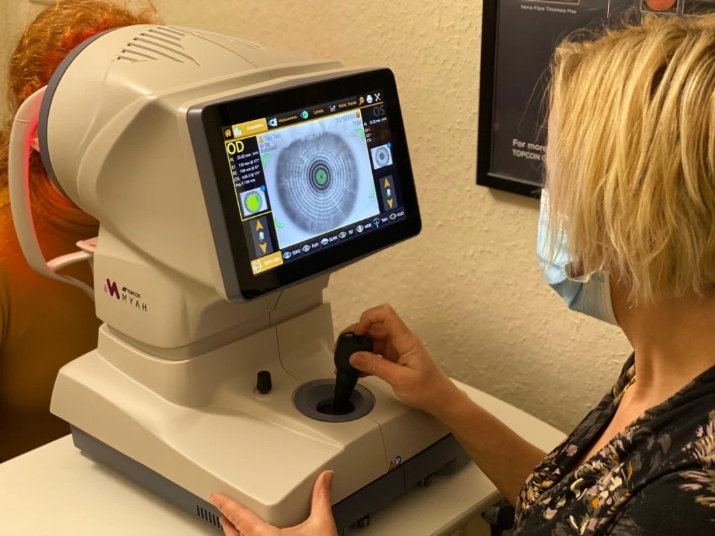
Corneal Topography Mapping Session
Following the initial examination, a corneal topography mapping session is scheduled. During this session, the corneal topographer captures detailed images of the cornea’s surface, providing precise measurements of its curvature and shape.
This data is crucial for designing contact lenses that fit the unique contours of the patient’s cornea, especially in cases of irregular corneas or post-surgical changes.
Lens Design and Fitting
With the corneal topography data in hand, the eye care professional proceeds to design the custom contact lenses. This process involves selecting the appropriate lens material, curvature, diameter, and power to correct the patient’s specific vision issues.
Once the lenses are manufactured, a fitting session is conducted to ensure that the lenses fit properly and provide the desired vision correction. Adjustments may be made based on the patient’s feedback and further assessments.
Follow-up Adjustments and Care Tips
After the initial fitting, follow-up appointments are crucial to monitor the lenses’ performance and the health of the patient’s eyes. These appointments allow the eye care professional to make any necessary adjustments to the lenses and provide guidance on proper lens care and handling.
Patients are also educated on recognizing signs of potential complications and advised on when to seek professional help.
Conclusion
Obtaining custom contact lenses is a detailed process that requires careful planning and execution. From the initial consultation to the follow-up adjustments, each step is designed to ensure that the lenses provide the best possible fit, comfort, and vision correction.
Corneal topography plays a pivotal role in this process, offering the detailed data needed to design lenses tailored to the unique needs of each patient’s eyes. By following these steps and maintaining open communication with their eye care professional, patients can achieve optimal results from their custom contact lenses.
VI. Final Note
The journey through understanding and utilizing corneal topography for specialized contact lens fittings underscores the significant advancements in eye care technology and personalized medicine. This sophisticated tool not only enhances the accuracy of contact lens fittings but also ensures that individuals with unique corneal conditions receive the best possible vision correction tailored to their specific needs.
Recap of the Importance of Precise Contact Lens Fitting
Corneal topography has proven to be an invaluable asset in the field of optometry and ophthalmology, particularly for those requiring specialized contact lenses. By providing a detailed map of the cornea’s surface, this technology allows for the design of lenses that fit precisely, thereby maximizing comfort and minimizing potential complications.
For patients with conditions like keratoconus, post-surgical corneal changes, or other irregularities, the use of corneal topography-guided lens fittings can dramatically improve their quality of life by enhancing visual clarity and reducing discomfort.
Encouragement for Seeking Expert Advice
For those experiencing discomfort with standard contact lenses or diagnosed with a corneal condition, it is crucial to seek the expertise of an eye care professional who specializes in custom contact lens fittings.
These specialists can provide comprehensive assessments using corneal topography to determine the most effective lens type and fit for each individual. Moreover, ongoing advancements in corneal topography technology continue to improve the precision and effectiveness of contact lens fittings, promising even better outcomes for patients in the future.
In conclusion, the integration of corneal topography into the process of contact lens fitting represents a significant step forward in personalized eye care. It empowers both patients and eye care professionals with the information needed to make informed decisions about eye health and vision correction.
As technology advances, the potential for even more precise and effective treatments continues to expand, offering hope and improved vision to those with complex corneal conditions.
This Article is Medically Reviewed by Oh Poh Ling

Poh Ling graduated as an optometrist from SEGi University. She believes that a person will be able to fully enjoy life when they have comfortable vision and healthy eyes. Poh Ling is involved in numerous vision screenings for the underprivileged school children and also for the public in an aim to promote awareness about the importance of regular eye examination. She enjoys travelling and playing tennis.
Her Specialties includes:
1. Specialty contact lens fitting: Keratoconus
2. Orthokeratology
Favourite Quote: “While there’s life, there is hope.” – Stephen Hawking